
 Every day, there are 79 organ transplants received by American patients as treatment for end-stage organ failure, according to statistics kept by the U.S. Department of Health & Human Services. However, the number of organs available for transplantation often doesn’t meet the need and an average of 22 U.S. citizens die every day from a lack of available organs for transplants. According to the American Transplant Foundation, the country’s waiting list for organ transplants has eclipsed 121,000 patients.
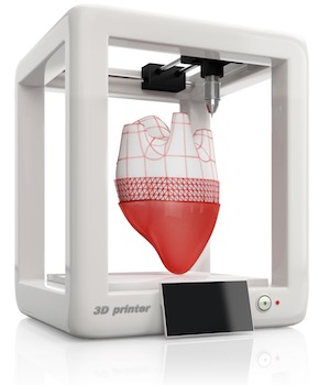
Given the fact that the country’s organ transplant waiting list has grown in volume by about 100,000 people since the early 1990s, any discovery that would increase the availability of transplantable organs would be a great benefit to society. In the middle of February, the scientific journal Nature Biotechnology published a paper from a team of researchers at Wake Forest University which reported a breakthrough in creating transplantable human organs with the use of an integrated tissue-organ printer. The team was successful in 3D printing an ear which survived on a mouse for two months and formed new tissues, indicating a successful integration to the new body.
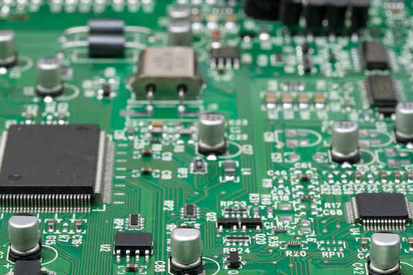
3D printing technologies have been a growing focus across the tech world as well as here on IPWatchdog, especially in recent months. Last year’s Consumer Electronics Show in Las Vegas showcased a wide range of applications for 3D printers, creating everything from clothing to circuit boards to candy. We’ve discussed how the growing trends in 3D printing, also known as additive manufacturing, could create a huge shift in industrial manufacturing by enabling solo innovators while also reducing waste seen in conventional manufacturing processes. Last October, IPWatchdog contributor Andrew Armstrong discussed the patentability of 3D printed organs in light of narrowed patentability for some biotechnologies under the America Invents Act.
Reports indicate that the total market for 3D printed body parts was $537 million in 2014 and that is likely to rise as artificial organs become more viable as a transplant option. Technological issues remain, however, in creating tissues and organs which are suitable for use in human transplants.
![[[Advertisement]]](https://ipwatchdog.com/wp-content/uploads/2024/04/Patent-Litigation-Masters-2024-banner-early-bird-ends-Apr-21-last-chance-938x313-1.jpeg)
The Wake Forest breakthrough provides 3D printed tissues with a biodegradable material to serve as a temporary framework for cells as they take hold in a host body; the tissue material also enables oxygen and nutrients to flow into the printed organ more easily. Still, issues in tissue complexity for certain organs remain.
Last year saw news of the world’s first 3D printed thyroid gland by a Russian firm called 3D Bioprinting Solutions. The firm unveiled the world’s first 3D printed thyroid gland, developed for use in treating a mouse with hypothyroidism, last March and reports indicate that the biotech firm was successful in transplanting that gland into a mouse by the end of the year. For the more than 20 million Americans who suffer from thyroid disorders, any steps towards the development of functional 3D printed structures that could serve as a replacement are helpful.
We’re also closer than ever to the world’s first 3D printed liver thanks in part to work performed by engineers at the University of California, San Diego. A paper published in early February by the online edition of Proceedings of the National Academy of Sciences discusses the scientist’s discoveries in creating tissues which closely reflect the structure and functioning of the liver, one of the body’s more complex organs. These liver tissues could help pharmaceutical companies developing liver treatments cut their costs in developing personalized treatments without the time-consuming and expensive need for clinical trials with humans or animals. The process involves the printing liver tissues from pluripotent cells collected from a patient’s skin, offering a minimally invasive way of collecting tissues that exhibit key liver functions, including urea production and albumin secretion.
For 3D printed organs to be able to take hold in a patient’s body and integrate into the body in a healthy way, the issue of how to provide nutrients from blood cells throughout a printed structure has proven to be a perplexing one. The recent Wake Forest advance addresses this problem by building micro-channels into the printed organs which allowed blood vessels to enter into the tissue material. Eventually, blood vessels were able to grow their way into the mouse’s ear and replace the micro-channels originally contained in the printed appendage.
The integrated tissue-organ printer, or ITOP, used by the Wake Forest team to print the artificial ear is capable of building synthetic tissues with living cells that are suspended within a polymer which is liquid but becomes gelatinous in consistency immediately after leaving the printer. The ITOP machine prints the polymer simultaneously with the biodegradable outer mold to create a structurally sound tissue lattice. Such ITOP printers can create highly individualized organs, if not yet incredibly complex ones, with patient data culled from magnetic resonance imaging (MRI) or computerized tomography (CT) scans. The printing process employed by the ITOP is more time-consuming than other 3D printing methods attempted for bodily organs but it’s able to produce larger viable tissues.
The use of a patient’s cells when bioprinting a transplantable organ also solves another issue which has plagued organ transplant procedures over the years: the rejection of an organ implant by a patient’s immune system. For instance, about 17 percent of kidney implants fail within three years after the transplant procedure, according to statistics reported by the National Kidney Foundation.
Such a system of 3D printing human tissues using a patient’s own cells is being pioneered by a team of researchers at Carnegie Mellon University. A study recently published by the research team in the scientific journal Science Advances describes the team’s development of the Freeform Reversible Embedding of Suspended Hydrogels (FRESH) method, a system in which soft materials such as collagen or fibrin are printed within a slurry of gelatin microparticles. Living cells can be added after the organ has been bioprinted with FRESH techniques and the gelatin melts away when the synthetic organ has been heated to 37°C (98.6°F), which is the normal temperature of the human body. Highly specialized bioprinters can cost tens of thousands of dollars, but the team from Carnegie Mellon was able to utilize open source software to implement FRESH on a consumer-level 3D printer which cost less than $1,000. Researchers believe that the technique could be used to create arteries, heart tissues or many other types of tissues or organs.
Although bones don’t have the same level of nutrient requirements of other bodily structures, there are still advances to be made in bioprinting bones, especially those protecting the most important nerve bundles in our bodies. Last December saw the world’s first successful surgery involving the implantation of a 3D printed vertebrae when an Australian neurosurgeon removed and replaced human vertebrae with 3D printed copies made from titanium. The 15-hour operation removed two vertebrae having cancerous tumors from the top of the patient’s spine. As of the middle of February, news reports indicate that the transplant has been successful, notwithstanding some unexpected complications in the patient’s eating and speaking as a result of the surgical procedure.
![[Advertisement]](https://ipwatchdog.com/wp-content/uploads/2024/04/Patent-Litigation-Masters-2024-sidebar-early-bird-ends-Apr-21-last-chance-700x500-1.jpg)

![[Advertisement]](https://ipwatchdog.com/wp-content/uploads/2021/12/WEBINAR-336-x-280-px.png)
![[Advertisement]](https://ipwatchdog.com/wp-content/uploads/2021/12/2021-Patent-Practice-on-Demand-recorded-Feb-2021-336-x-280.jpg)
![[Advertisement]](https://ipwatchdog.com/wp-content/uploads/2021/12/Ad-4-The-Invent-Patent-System™.png)


Join the Discussion
No comments yet.